Calcaneal Locking Plate
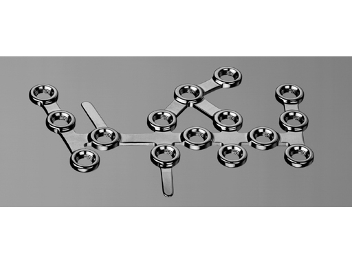
The Calcaneal Locking Plates are designed to address complex fractures and osteotomies of the calcaneus, including, but not limited to extraarticular, intraarticular, joint depression, tongue type and severely comminuted fractures. The plates are applied to the lateral side. The plate has 15 threaded holes, which accept 2.7 mm and 3.5 mm Cortex Screws as well as 3.5 mm Locking Screws. The numerous holes enable versatility and provide options to address multiple fragments and fracture patterns. It provides a fixed angle construct and a buttress to the articular surface of the calcaneus. The distal end of the plate has two bendable tabs to give support for the anterior process and plantar fragments. The plate is available in two sizes, short and large, and comes in a left and a right version. At the moment, the plate is available in stainless steel. A version in a special kind of titanium with 15% molybdenum is under development. Several new instruments were developed for the convenient use of this plate:
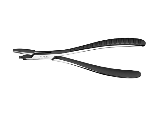
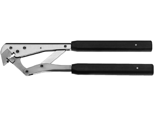
Tab Bender Pliers for Calcaneal Locking Plate
The Tab Bending Pliers are used to bend the two taps located on the Calcaneal Locking Plates. The surgeons now have the ability to safely contour the tabs of the plate in-situ for better anatomical fit.
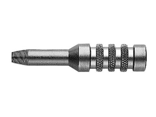
2.8 mm Threaded Drill Guide
Guides a 2.8 mm drill bit perpendicular to the plate to ensure proper mating of the 3.5 mm Locking Screw and plate hole threads.
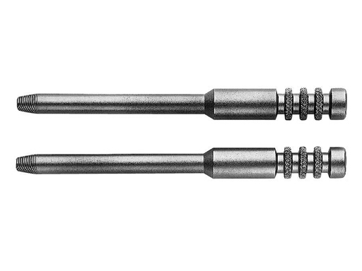
Threaded Plate Holders
Aids to position the plate to the bone and may also be used for in-situ bending to achieve a better plate fit.
Calcaneal Locking Plate Cutter
Facilitates precise hole/tab removal without creating upward facing burrs.
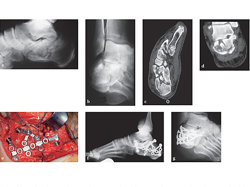
First clinical use (January 4, 2000, Dresden) of the new interlocking calcaneal plate in a B2 fracture of the calcaneus
The lateral view and the Brodns view show the deep impaction of the posterior facet (see Fig a-b).
The axial and coronal cut of the CT scan show the severe damage to the calcaneo-cuboidal and subtalar joint classified according to the new AO-Classification as 81.2 B2 [ie, (1.3.3)]. 81.2 = calcaneus, B2 = intraarticular fracture of two joints involved, d = posterior facet, h = cuboidal facet, 1.3.3 = bony lesion, multifragmentary, severely distocated (see Fig c-d).
Intraoperative situation after anatomical reduction and fixation with the interlocking plate. 1 = one of the four 2.0 mm K-wires, which are inserted in the talus and cuboid keeping the full thickness flap upwards for optimal exposure. 2 = tab bowed close to the bone in the calcaneal neck area to keep the ante (see Fig e-g).
Hazards and labeling
Due to varying countries’ legal and regulatory approval requirements, consult the appropriate local product labeling for approved intended use of the products described on this website. All devices on this website are approved by the AO Technical Commission. For logistical reasons, these devices may not be available in all countries worldwide at the date of publication.
Legal restrictions
This work was produced by AO Foundation, Switzerland. All rights reserved by AO Foundation. This publication, including all parts thereof, is legally protected by copyright.
Any use, exploitation or commercialization outside the narrow limits set forth by copyright legislation and the restrictions on use laid out below, without the publisher‘s consent, is illegal and liable to prosecution. This applies in particular to photostat reproduction, copying, scanning or duplication of any kind, translation, preparation of microfilms, electronic data processing, and storage such as making this publication available on Intranet or Internet.
Some of the products, names, instruments, treatments, logos, designs, etc referred to in this publication are also protected by patents, trademarks or by other intellectual property protection laws (eg, “AO” and the AO logo are subject to trademark applications/registrations) even though specific reference to this fact is not always made in the text. Therefore, the appearance of a name, instrument, etc without designation as proprietary is not to be construed as a representation by the publisher that it is in the public domain.
Restrictions on use: The rightful owner of an authorized copy of this work may use it for educational and research purposes only. Single images or illustrations may be copied for research or educational purposes only. The images or illustrations may not be altered in any way and need to carry the following statement of origin “Copyright by AO Foundation, Switzerland”.
Check www.aofoundation.org/disclaimer for more information.
If you have any comments or questions on the articles or the new devices, please do not hesitate to contact us.
“approved by AO Technical Commission” and “approved by AO Foundation”
The brands and labels “approved by AO Technical Commission” and “approved by AO Foundation”, particularly "AO" and the AO logo, are AO Foundation's intellectual property and subject to trademark applications and registrations, respectively. The use of these brands and labels is regulated by licensing agreements between AO Foundation and the producers of innovation products obliged to use such labels to declare the products as AO Technical Commission or AO Foundation approved solutions. Any unauthorized or inadequate use of these trademarks may be subject to legal action.
AO ITC Innovations Magazine
Find all issues of the AO ITC Innovations Magazine for download here.
Innovation Awards
Recognizing outstanding achievements in development and fostering excellence in surgical innovation.


