2.4 mm VA-LCP Volar Rim Distal Radius Plate
Very distal, mostly intraarticular fractures of the distal radius may require fixation very close to the volar articular lip. Proper placement of standard plates is difficult due to variances of the anatomical shape of the bone and the origins of the radiocarpal ligaments which must not be injured. Moreover, fixation options of the radial styloid and distal radioulnar joint have not been optimal so far.
The 2.4 mm VA-LCP volar rim distal radius derives from the 2.4 mm LCP distal radius plate juxta-articular, but has an anatomically preshaped design, a second distal screw row (outrigger), and variable angle locking technology. Highly polished low profile plates with round edges and fully countersunk screws minimize the risk of softtissue irritations and tendon ruptures.
Fig 1a Volar rim distal radius plate.
The anatomical plate design allows for very distal plate placement. CT scans were used to verify the fit of the precontoured plate. Bendable outriggers aid in adjusting the precontoured plate to specific anatomical need and individual variations.
The second distal screw row provides for superior fixation stability of fragments, eg, radial styloid, and especially the most ulnar corner of the lunate fossa.
Fig 1b Volar rim distal radius plate.
Nonclinical dynamic-fatigue testing to determine fatigue strength of the plate construct showed that the VA plates are stronger than the 2.4 mm LCP distal radius plate juxta-articular.
The plate is available in stainless steel and titanium (TiCp), each in four different versions: 5-hole shaft with 6-hole head in left and right versions, and 5-hole shaft with 7-hole head in left and right versions. For ease and expedience each plate version has a specific guiding block for standard screw orientation. Trial implants help determine the correct plate dimension for sterile implant use.
Fig 2a-b Comparison of anatomical bend and screw placement of volar rim DR plate (a) and DR juxta-articular plate (b).
A 74-year-old woman sustained an intraarticular distal forearm fracture of the radius and ulna after falling on her outstretched hand.
Case provided by Daniel Rikli, Basel, Switzerland
Fig 1ab Preoperative x-rays.
Fig 2ab Initial management was with closed reduction and joint bridging external fixator. There was loss of primary reduction due to inadequate positioning of the external fixator (too lateral).
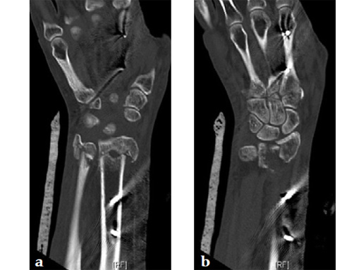
Fig 3ce Radius with hyperextended palmar-ulnar key fragment.
Fig 3f Ulnar head split, displaced dorsally (CT sagittal reconstruction).
Fig 4ab X-rays 3 days postoperatively: removable splint, early motion with physiotherapy.
Fig 5ab Six weeks postoperatively: residual swelling, little pain, range of motion improving with physiotherapy; the patient uses her hand for daily activities.
Hazards and labeling
Due to varying countries’ legal and regulatory approval requirements, consult the appropriate local product labeling for approved intended use of the products described on this website. All devices on this website are approved by the AO Technical Commission. For logistical reasons, these devices may not be available in all countries worldwide at the date of publication.
Legal restrictions
This work was produced by AO Foundation, Switzerland. All rights reserved by AO Foundation. This publication, including all parts thereof, is legally protected by copyright.
Any use, exploitation or commercialization outside the narrow limits set forth by copyright legislation and the restrictions on use laid out below, without the publisher‘s consent, is illegal and liable to prosecution. This applies in particular to photostat reproduction, copying, scanning or duplication of any kind, translation, preparation of microfilms, electronic data processing, and storage such as making this publication available on Intranet or Internet.
Some of the products, names, instruments, treatments, logos, designs, etc referred to in this publication are also protected by patents, trademarks or by other intellectual property protection laws (eg, “AO” and the AO logo are subject to trademark applications/registrations) even though specific reference to this fact is not always made in the text. Therefore, the appearance of a name, instrument, etc without designation as proprietary is not to be construed as a representation by the publisher that it is in the public domain.
Restrictions on use: The rightful owner of an authorized copy of this work may use it for educational and research purposes only. Single images or illustrations may be copied for research or educational purposes only. The images or illustrations may not be altered in any way and need to carry the following statement of origin “Copyright by AO Foundation, Switzerland”.
Check www.aofoundation.org/disclaimer for more information.
If you have any comments or questions on the articles or the new devices, please do not hesitate to contact us.
“approved by AO Technical Commission” and “approved by AO Foundation”
The brands and labels “approved by AO Technical Commission” and “approved by AO Foundation”, particularly "AO" and the AO logo, are AO Foundation's intellectual property and subject to trademark applications and registrations, respectively. The use of these brands and labels is regulated by licensing agreements between AO Foundation and the producers of innovation products obliged to use such labels to declare the products as AO Technical Commission or AO Foundation approved solutions. Any unauthorized or inadequate use of these trademarks may be subject to legal action.
AO ITC Innovations Magazine
Find all issues of the AO ITC Innovations Magazine for download here.
Innovation Awards
Recognizing outstanding achievements in development and fostering excellence in surgical innovation.


